Leg Bones Diagram : 1. The bones of the leg are the femur, tibia, fibula and patella. The femur, or thighbone, is the longest and largest bone in the human body. The larger of the two is the tibia, familiarly called the shinbone. The nerves of the leg and foot arise from spinal nerves connected to the spinal cord in the lower back and pelvis. How to draw hand bone diagram| phalange carpal bone diagram.
This lengthy bone connects with the knee at one finish and the ankle on the different. Tibia is a latin word meaning both shinbone and flute. The elbow is located below the chest at the back of the foreleg. In fact nearly one quarter of the bones in the body are found in the feet. It is thought that tibia refers to both the bone and the musical instrument because flutes were once fashioned from the tibia (of animals).
Labeled 3d Medical Illustration Of Male Pelvis Hip And Leg Bones On White Background Stock Photo Alamy from c8.alamy.com Related posts of diagram of leg bones inside of arm muscle and bone. The tibia and fibula which are the leg bones between the knee and ankle. Inside of arm muscle and bone 12 photos of the inside of arm muscle and bone , bone Its lower end helps create the knee joint. The tibia and the fibula. The femur, or thighbone, is the longest and largest bone in the human body. Now i will provide you the few information on the other bones of dog leg anatomy with their unique features. Subsequent to the tibia is the fibula, the thinner, weaker bone of the decrease leg.
The forearm is the long bone that runs just after the elbow.
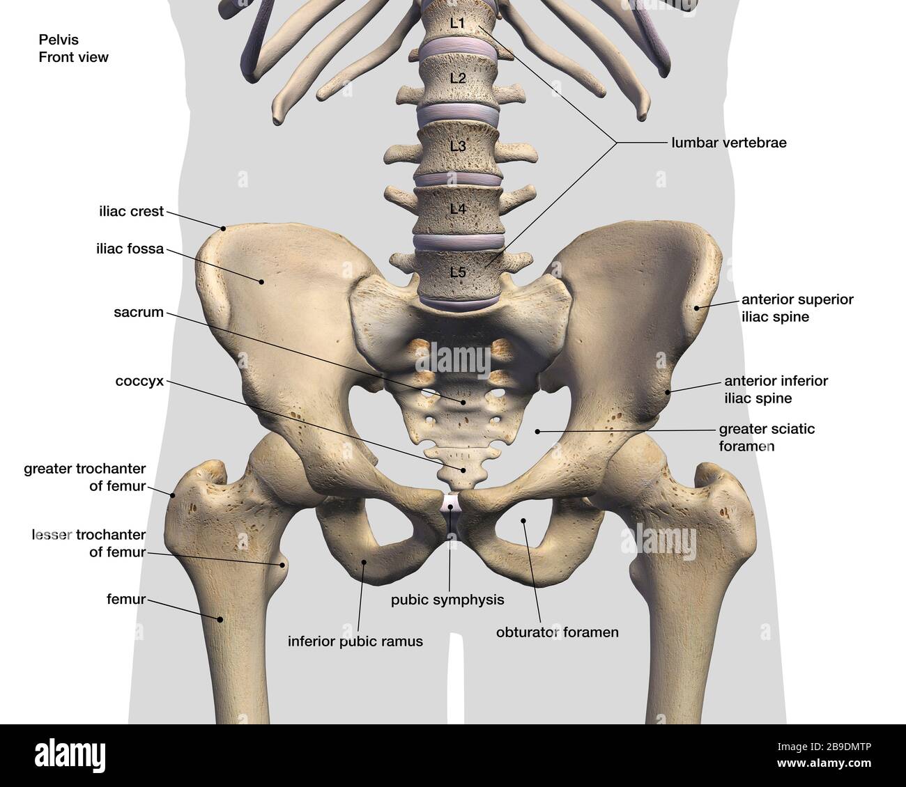
The smaller lateral bone of the lower leg. The tibia and the fibula. The tibia and the fibula, at the top of the ankle joint. There are three hamstring muscles, all of them originating at the ischial tuberosity (the bones you sit on): Ulna and the radius are two bones that sit next to each other. 1 and the one behind the little toe is no. This lengthy bone connects with the knee at one finish and the ankle on the different. The lower leg extends from the knee to the ankle. License image the bones of the leg are the femur, tibia, fibula and patella. The bones of the hip include the femur, the ilium, the ischium, and the pubis. Human anatomy diagrams show internal organs, cells, systems, conditions, symptoms and sickness information and/or tips for healthy living. They support the legs to bear the body weight and also help in proper locomotion. The bones of the leg are the femur, tibia, fibula and patella.the foot bones shown in this diagram are the talus, navicular, cuneiform, cuboid, metatarsals and calcaneus.
With different grades of sprains depending on severity. The bones of the hip include the femur, the ilium, the ischium, and the pubis. In fact nearly one quarter of the bones in the body are found in the feet. Related posts of diagram of leg bones inside of arm muscle and bone. The lower leg extends from the knee to the ankle.
18 464 Leg Bone Stock Photos Pictures Royalty Free Images Istock from media.istockphoto.com Anchor chart diagram leg human knee skeleton health bone science human body. The pubis, ischium, and ilium together constitute the pelvis while the thigh bone is the femur. The bones together make up the hip. The lower leg is comprised of two bones, the tibia and the smaller fibula. The bones of the leg and foot form part of the appendicular skeleton that supports the many muscles of the lower limbs. How to draw a human bone leg. The forearm is the long bone that runs just after the elbow. Its lower end helps create the knee joint.
The proximal portion of the tibia is tibial plateau which acts as a cusp for the knee, the distal portion tapers into the medial malleoli and the concave surface which articulates with the talus at the ankle joint.
The lower leg extends from the knee to the ankle. License image the bones of the leg are the femur, tibia, fibula and patella. The larger of the two is the tibia, familiarly called the shinbone. See more ideas about muscle anatomy, human anatomy and physiology, medical anatomy. The elbow is located below the chest at the back of the foreleg. The femur, or thighbone, is the longest and largest bone in the human body. The nerves of the leg and foot arise from spinal nerves connected to the spinal cord in the lower back and pelvis. The axial skeleton, comprising the spine, chest and head, contains 80 bones. Now i will provide you the few information on the other bones of dog leg anatomy with their unique features. He leg's main function in the human is for locomotion and support of the rest of the. Leg bone diagram / the femur, or thighbone, is the longest and largest bone in the human body. Click now to learn more about the bones, muscles, and soft tissues tibia: The human leg consists of 8 bones, 4 per leg.
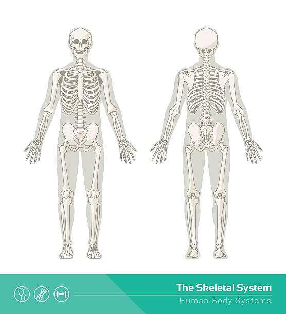
Also called the shin bone, the tibia is the longer of the two bones in the. The femur, or thighbone, is the longest and largest bone in the human body. The elbow is located below the chest at the back of the foreleg. The leg has two bones: The appendicular skeleton, comprising the arms and legs, including the shoulder and pelvic girdles, contains 126 bones, bringing the total for the entire skeleton to 206 bones.
Bones Of The Lower Limb Anatomy And Physiology from s3-us-west-2.amazonaws.com Related posts of leg bones anatomy diagram cross section of foot nerves. These muscles work together to produce movements such as standing, walking, running, and jumping. 1 and the one behind the little toe is no. How to draw hand bone diagram| phalange carpal bone diagram. This area is commonly referred to as the calf. Top suggestions for human leg bones diagram. Use lucidchart to visualize ideas, make charts, diagrams & more. The bones together make up the hip.
Numerous bone is the long bone of the upper arm which goes all the way to the elbow.
Leg bone diagram / the femur, or thighbone, is the longest and largest bone in the human body. Inside of arm muscle and bone 12 photos of the inside of arm muscle and bone , bone How to draw a human bone leg. Tibia and fibula the tibia and fibula are two long bones that run parallel to each other, forming the scaffold of the leg and providing attachment points for many muscles. At the same time, the bones and joints of the leg and foot must be strong enough to support the body's weight while remaining. Numerous bone is the long bone of the upper arm which goes all the way to the elbow. Below given knee diagram will help you to understand the various parts and functioning of the knee joint. Bone diagram forehead (frontal bone) nose bones (nasals) cheek bone (zygoma) upper jaw (maxilla) lower jaw (mandible) breast bone (sternum) upper arm bone (humerus) lower arm bone (ulna) thigh bone (femur) collar bone (clavicle) toe bones (phalanges) ankle bones (tarsals) kneecap (patella) shin bone Human anatomy diagrams show internal organs, cells, systems, conditions, symptoms and sickness information and/or tips for healthy living. Ulna and the radius are two bones that sit next to each other. The bones together make up the hip. He leg's main function in the human is for locomotion and support of the rest of the. In fact nearly one quarter of the bones in the body are found in the feet.


0 Response to "Leg Bones Diagram : 1"
Post a Comment